Conditions
Submucosal Tumours of the Stomach and Oesophagus (GIST, NET, Leiomyoma, and Schwannoma)
What is it?
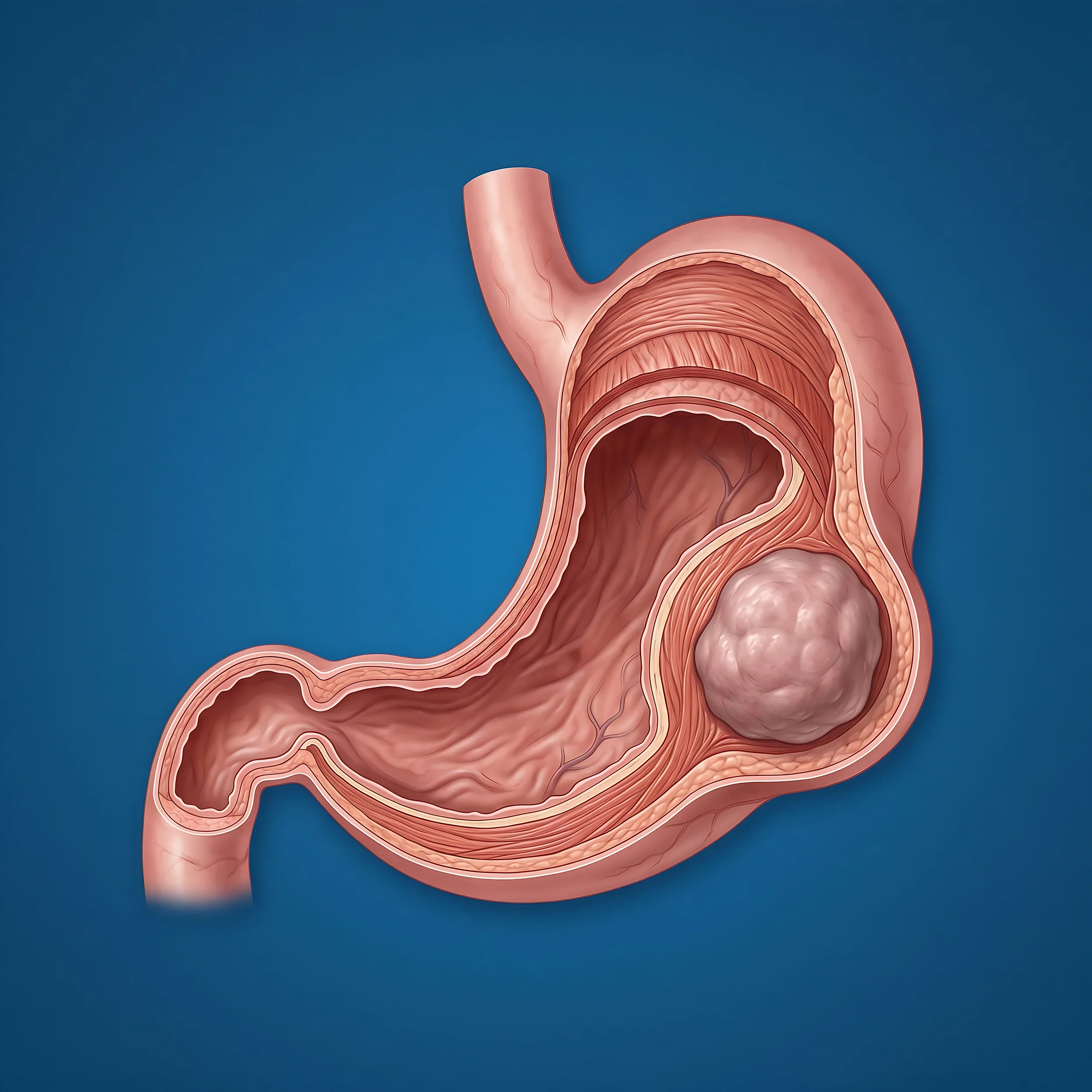
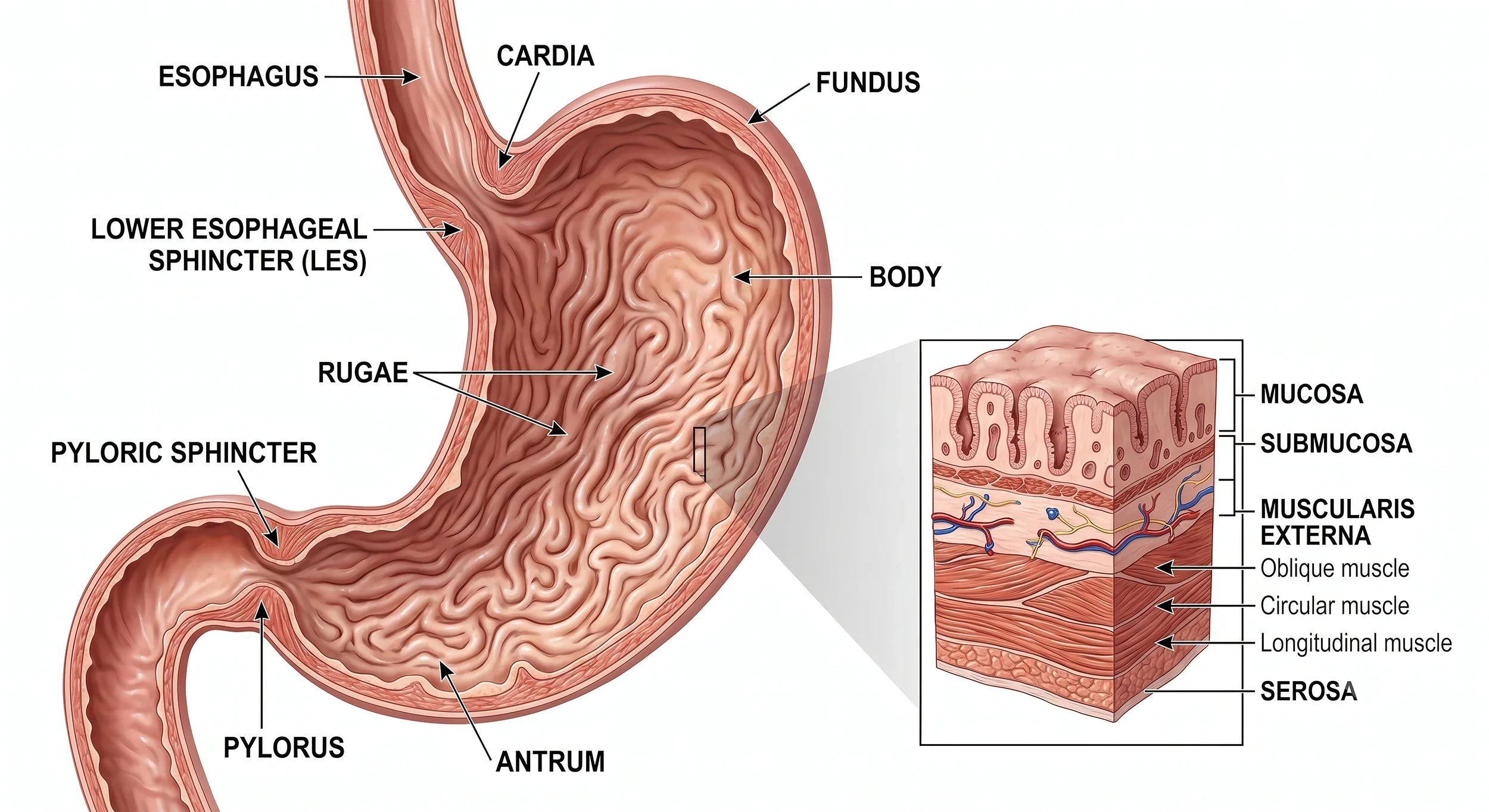
Most cancers of the stomach and oesophagus start in the lining (mucosa) that food passes over. Submucosal tumours are different — they arise from the layers underneath the lining, such as muscle, nerve, or hormone-producing tissue. Because they grow beneath the surface, the lining itself often looks normal on endoscopy, with the tumour appearing as a smooth bulge underneath it.
There are several types, each arising from a different tissue:
GIST (gastrointestinal stromal tumour) — arises from specialised cells in the wall of the gut that help control its movement. GISTs can behave in different ways: some stay small and low-risk, while others have the potential to grow or spread if left untreated.
NET (neuroendocrine tumour) — arises from hormone-producing cells within the gut wall. These also vary in behaviour, from slow-growing and low-risk to more active types requiring closer management.
Leiomyoma — a benign (non-cancerous) tumour of smooth muscle.
Schwannoma — a benign tumour arising from the cells that form the sheath around nerves.
Leiomyomas and schwannomas are almost always benign. GISTs and NETs can occasionally behave more aggressively, which is why they're assessed more carefully once found.
How are they usually found?
Most submucosal tumours cause no symptoms at all and are found incidentally — for example, during an endoscopy or CT scan performed for an unrelated reason.
What symptoms should you look out for?
When symptoms do occur, they depend on the size and location of the tumour, and can include:
Vomiting blood, or vomit or stool that looks like coffee grounds or is black and tarry
Unexplained anaemia (low blood count), sometimes picked up on routine blood tests
Abdominal discomfort or a feeling of fullness
Difficulty swallowing, if the tumour is in the oesophagus
Occasionally, a lump that can be felt
How are they diagnosed and investigated?
Endoscopy — usually the first test, allowing the tumour to be seen and its size and location assessed.
Endoscopic ultrasound (EUS) — a specialised camera test combining endoscopy with ultrasound, used to look closely at which layer of the wall the tumour arises from and its features. A fine needle biopsy can often be taken at the same time to get a tissue diagnosis.
CT scan — used to assess the size and extent of the tumour, and to check there's no spread elsewhere.
Depending on the type of tumour suspected, additional specialised tests may be arranged, particularly for NETs, to build a complete picture before deciding on treatment.
Does every submucosal tumour need treatment?
Not necessarily. Many small, low-risk tumours — particularly leiomyomas, schwannomas, and small GISTs or NETs without any concerning features — can simply be monitored with periodic endoscopy or scans, rather than removed. Surgery tends to be recommended when a tumour is growing, causing symptoms, has features suggesting higher risk, or when a firm diagnosis can't be reached without removing it.
What are the benefits of surgery?
Removes the tumour completely — eliminating any risk of future growth or change in behaviour.
Provides a definitive diagnosis — examining the whole tumour under the microscope gives certainty about exactly what it is, which isn't always possible from a needle biopsy alone.
Can be curative — for tumours confined to the wall of the stomach or oesophagus, surgical removal is often all that's needed.
What are the alternatives to surgery?
Surveillance — regular endoscopy, EUS, or CT scans to monitor the tumour over time, reserving surgery for if it changes.
Targeted drug therapy — for some GISTs, particularly larger or higher-risk ones, medication (such as imatinib, brand name Glivec) can shrink the tumour before surgery or control it instead of surgery, depending on the situation.
No treatment — reasonable for some small, incidental, low-risk findings, particularly leiomyomas and schwannomas.
I will discuss which of these options suits your particular tumour and situation.
What does the surgery involve?
The type of operation depends on the size, location, and type of tumour:
Local excision (wedge resection) — removing the tumour along with a margin of normal surrounding tissue, most often performed laparoscopically (keyhole surgery) for stomach tumours. This is the most common approach for GISTs, leiomyomas, and schwannomas.
Endoscopic removal — in select cases of small, superficial tumours, removal can sometimes be achieved during endoscopy without the need for an abdominal operation.
More extensive resection — occasionally, if a tumour is large or in a difficult location, a bigger operation (such as a partial gastrectomy or oesophagectomy) may be needed instead.
Unlike typical stomach or oesophageal cancers, removal of surrounding lymph nodes usually isn't necessary for GISTs, leiomyomas, or schwannomas, as they don't tend to spread that way. NETs are assessed individually, as this can vary.
What are the risks of surgery?
As with any operation, there are risks. I will discuss these with you in more detail, but in general terms:
Common, usually minor — wound discomfort and temporary adjustment to eating while your digestive system settles.
Less common — chest infection, wound infection, or a small collection of fluid near the surgical site.
Uncommon — leakage from the staple line or join, or bleeding.
Rare — injury to nearby structures, or other more serious complications requiring further treatment.
Most people recover from this surgery without a serious complication, and the risks are generally lower than for the more extensive cancer operations, given that these tumours are usually removed with a smaller, more localised procedure.
What does recovery look like?
Hospital stay — typically a few days for a local excision, though this can be longer if a more extensive operation is required.
Eating and drinking — most people can start on fluids soon after surgery and build back up to a normal diet over a short period.
Return to normal activity — recovery from a local excision is usually quicker than from a major cancer resection, though this varies with the extent of surgery performed.
Follow-up — some tumours, particularly GISTs and NETs, require ongoing surveillance with scans after removal, even once successfully treated, to ensure continued wellbeing. This will be tailored to your specific diagnosis.
Summary
Submucosal tumours (GIST, NET, leiomyoma, and schwannoma) arise from the deeper layers of the stomach or oesophageal wall, rather than the lining itself.
Most are found incidentally and cause no symptoms; some can cause bleeding, anaemia, or discomfort.
Endoscopic ultrasound and biopsy, along with CT scanning, are the key tests used to characterise these tumours.
Not all require surgery — some are simply monitored, while others benefit from removal.
Surgery, usually a local excision, gives a definitive diagnosis and can be curative, with recovery generally quicker than for major cancer surgery.
This information is general in nature and is not intended as a substitute for a full discussion of the risks and benefits of surgery considering your medical issues. If you have further questions or have been diagnosed with a submucosal tumour, please make an appointment.
Download: Partial Gastrectomy/GIST Post-Op Pack
Next Step
A referral is required prior to your appointment. Call (03) 9923 8066 or email admin@shilton.net.au to arrange a consultation.